Chiari Malformation
Diagnosis and decompression surgery for Chiari malformation in Lahore, Pakistan
Found Chiari Malformation on an MRI Done for Headaches? What It Means
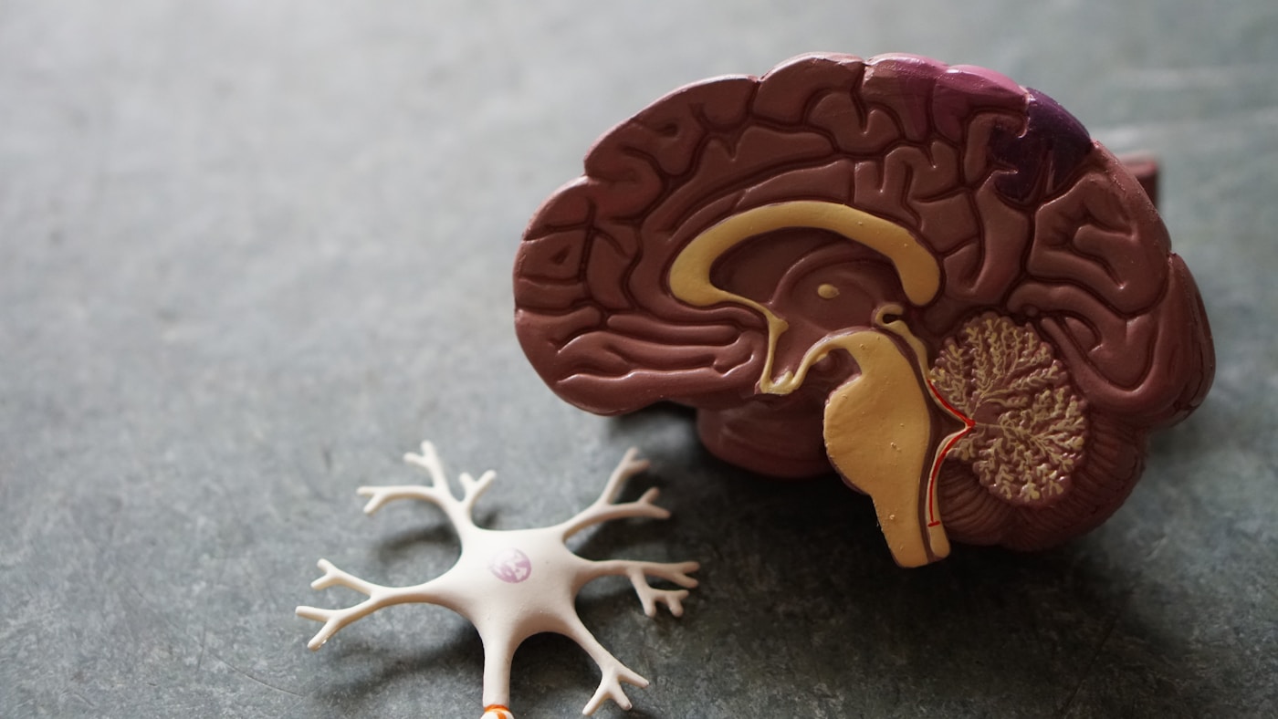
A Chiari malformation (also called Arnold-Chiari malformation) is a structural problem at the base of the skull, in which the cerebellar tonsils — the lowest part of the cerebellum — extend down through the foramen magnum (the natural opening at the base of the skull) into the upper part of the spinal canal.
It is very common for a Chiari malformation to be picked up unexpectedly — a patient has an MRI of the brain done because of recurring headaches, neck pain, or dizziness, and the radiology report mentions "cerebellar tonsillar descent" or "low-lying cerebellar tonsils." Understandably, this can be alarming to read. In reality, a large proportion of these findings are mild, incidental, and do not need surgery — but the finding does need to be reviewed by a neurosurgeon alongside your actual symptoms, because the significance of a Chiari malformation depends heavily on how far the tonsils descend, whether there is any crowding of the brainstem, and whether a syrinx (fluid cavity) has formed in the spinal cord.
This crowding at the base of the skull can compress the brainstem and upper spinal cord, and can also block the normal flow of cerebrospinal fluid (CSF) between the brain and spinal cord. Dr. Waqas Mehdi, FCPS Neurosurgeon at MidCity Hospital Lahore, evaluates patients with Chiari malformation — many of whom are found incidentally on an MRI done for headaches or neck pain — and advises clearly on whether observation or decompression surgery is the right course of action.
Symptoms of Chiari Malformation
Why Does a Chiari Headache Get Worse with Coughing or Straining?
One of the most recognisable features of a Chiari-related headache is that it is triggered or sharply worsened by coughing, sneezing, laughing, lifting something heavy, or straining during a bowel movement — actions that all involve what is known as the Valsalva maneuver.
During a Valsalva maneuver, pressure rises suddenly inside the chest and abdomen, and this pressure is transmitted upward into the veins and cerebrospinal fluid space around the spinal cord and brain. In a normal foramen magnum, there is enough room to absorb this momentary pressure surge. But when the cerebellar tonsils are already crowding the foramen magnum, this extra pressure has nowhere to go — it pushes the already-crowded tissue further against the brainstem and briefly worsens the obstruction to CSF flow, producing a sudden, sharp, occipital (back-of-head) headache that can last seconds to a few minutes and then ease off.
This specific pattern — a headache tied tightly to coughing, straining, or bending forward, felt at the back of the head or upper neck — is one of the most useful clues that points a doctor toward a Chiari malformation rather than a more common headache disorder like tension headache or migraine, and is one of the first questions Dr. Waqas Mehdi asks when reviewing a patient with an incidental Chiari finding.
Chiari I vs Chiari II vs Chiari III — Understanding the Types
Chiari Type I
By far the most common type, and the one most often found incidentally in teenagers and adults during an MRI done for headaches, neck pain, or unrelated reasons. The cerebellar tonsils descend below the foramen magnum, usually by a small amount. Many Type I cases are mild and asymptomatic; others cause the classic occipital headache, neck pain, or syringomyelia and need decompression surgery.
Chiari Type II
A more significant malformation, almost always diagnosed in infancy, and strongly associated with spina bifida (myelomeningocele) and hydrocephalus. Along with the cerebellar tonsils, the brainstem and fourth ventricle are also displaced downward. This type is typically identified on prenatal or newborn imaging and is managed jointly with pediatric neurosurgery.
Chiari Type III
A rare and severe form in which part of the cerebellum and brainstem herniate through a defect at the back of the skull or upper neck into a sac (an occipital or high cervical encephalocele). This requires early, specialised neurosurgical management, usually in infancy.
Chiari Type IV
The rarest and most severe type, involving underdevelopment (hypoplasia) of the cerebellum itself rather than simple displacement. This type carries a guarded outlook and is diagnosed very early in life.
Most patients researching this topic after an adult MRI finding have Chiari Type I — the mildest and most manageable of the four types.
What Is Syringomyelia and How Is It Connected to Chiari?
Syringomyelia refers to a fluid-filled cavity, called a syrinx, that forms within the spinal cord itself. It is one of the most important things a neurosurgeon looks for on the MRI of anyone diagnosed with a Chiari malformation, because it changes both the urgency and the recommended treatment.
When the cerebellar tonsils crowd the foramen magnum in a Chiari malformation, normal cerebrospinal fluid flow between the brain and spinal cord is disrupted. Over time, this abnormal flow pattern is believed to force fluid into the central canal of the spinal cord, gradually expanding it into a syrinx. As the syrinx grows, it can damage the nerve fibres that carry pain and temperature sensation across the middle of the spinal cord, which is why patients with syringomyelia often notice a curious pattern — loss of ability to feel pain or temperature (for example, not noticing a hot cup of tea) in the hands and arms, while normal touch sensation is preserved.
If left untreated for a long time, a syrinx can slowly enlarge and cause progressive weakness, loss of hand function, or worsening sensory loss. The encouraging part is that syringomyelia caused by a Chiari malformation is usually a secondary, downstream effect of the crowding at the foramen magnum — treating the underlying cause with posterior fossa decompression surgery restores more normal CSF flow, and in most cases the syrinx gradually shrinks or stabilises over the following months, tracked with follow-up MRI. Separate surgery on the syrinx itself is rarely needed once the underlying Chiari malformation has been properly decompressed.
Chiari Malformation in Children vs Adults
In Children
Chiari II is usually diagnosed early because of its strong link with spina bifida and hydrocephalus, and is managed as part of a coordinated pediatric neurosurgical plan from infancy. Chiari I in children can present with headaches, scoliosis (curvature of the spine), swallowing difficulty, or sleep-disordered breathing, and sometimes with developmental or behavioural changes that are easy to miss if no one is specifically looking for a Chiari malformation.
In Adults
Chiari I is the type almost always seen in adults, and a large share of cases are picked up incidentally on MRI done for headache or neck pain rather than because of dramatic symptoms. Adult presentations tend to centre on the classic straining-related occipital headache, neck pain, dizziness, and — if a syrinx has developed — subtle hand numbness or weakness that may have been present for years before diagnosis.
Diagnosis
Does Chiari Malformation Always Need Surgery?
No — this is one of the most common and important questions asked after an incidental Chiari finding. A significant number of people, particularly those with a small degree of tonsillar descent and no syrinx, have no symptoms at all and will never need any treatment beyond periodic monitoring.
Surgery is generally considered when there are clear symptoms that match the MRI findings (particularly the characteristic straining-related occipital headache), when there is a syrinx in the spinal cord, or when the CINE MRI flow study shows significant obstruction of CSF flow at the foramen magnum. The decision is individualised — it is never made from the MRI report alone, but from a combination of imaging, CSF flow assessment, and a detailed clinical picture.
Treatment Options
Observation
For patients with a mild, incidentally discovered Chiari malformation and no significant symptoms, periodic clinical review and MRI monitoring is often all that is needed. Most such patients live a normal life without ever requiring surgery.
Posterior Fossa Decompression Surgery — What Is Actually Done
The standard surgical treatment for symptomatic Chiari malformation. Under general anaesthesia, a small area of bone is removed at the back of the skull (suboccipital craniectomy) and, when needed, the back arch of the first cervical vertebra (C1 laminectomy) to relieve crowding at the foramen magnum. In many cases the dura — the tough membrane covering the brain and spinal cord — is opened and enlarged with a patch (duraplasty) to give the cerebellum and CSF pathways more room and restore more normal fluid flow.
Hospital Stay and Recovery
Patients are typically monitored in hospital for a few days after surgery. Neck stiffness and mild headache in the first one to two weeks are common and expected as the tissues heal. Most patients gradually return to normal daily activities over four to six weeks, with heavy lifting and strenuous activity restricted for a longer period on the surgeon's advice. Headache and other symptoms often begin to improve within the first few weeks, though the timeline varies from patient to patient, and any accompanying syrinx typically takes longer — months — to shrink on follow-up MRI.
Management of Associated Syringomyelia
In most cases, successful decompression surgery allows the syrinx to shrink over time without needing separate treatment. Follow-up MRI at intervals tracks this response and confirms the CSF pathways have been adequately restored.
Symptomatic Treatment
Pain management and physiotherapy can help with headache and neck symptoms in patients being observed or recovering from surgery.
Frequently Asked Questions
What is a Chiari malformation?
Who treats Chiari malformation in Lahore?
What are the symptoms of Chiari malformation?
Does Chiari malformation always need surgery?
What does Chiari decompression surgery involve?
Is Chiari malformation life-threatening?
Can Chiari malformation cause anxiety or dizziness?
How is Chiari malformation different from a brain tumor?
What is the success rate of Chiari decompression surgery?
Can Chiari malformation be hereditary?
Found to Have a Chiari Malformation on MRI?
Consult Dr. Waqas Mehdi for a clear explanation of what your MRI shows and whether treatment is needed. MidCity Hospital, Lahore — Mon to Sat, 6–8 PM.
Related Conditions & Services
Consult Dr. Waqas Mehdi Today
Pakistan's trusted neurosurgeon for brain tumor surgery, spine surgery, and all complex neurological conditions. Book your appointment at MidCity Hospital, Lahore.