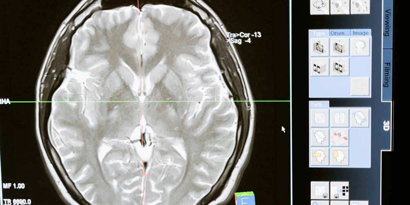
Hydrocephalus (Water on the Brain)
Understanding hydrocephalus — causes, symptoms, and surgical treatment options available in Lahore, Pakistan
What is Hydrocephalus?
Hydrocephalus is a build-up of cerebrospinal fluid (CSF) inside the cavities (ventricles) of the brain. CSF is normally produced, circulated, and reabsorbed in a continuous cycle; when this cycle is disrupted — either because fluid cannot drain properly or is produced faster than it is absorbed — it accumulates and increases pressure inside the skull. The word hydrocephalus comes from the Greek words for "water" and "brain."
In Pakistan, hydrocephalus is commonly seen in newborns and infants due to congenital causes, as well as in children following meningitis, intraventricular hemorrhage of prematurity, or head injury. Adults can develop hydrocephalus from brain tumors, bleeding, infection, or the aging process (Normal Pressure Hydrocephalus). The right treatment and long-term outlook depend heavily on which of these patterns applies — which is why an accurate diagnosis matters as much as the surgery itself.
Is My Baby's Head Growing Too Fast? Warning Signs Parents Miss
Every baby's head grows in the first year of life, which is exactly why abnormal growth is often missed — parents and even some clinicians assume a large head is simply the child's build. The key difference is the rate of growth: in hydrocephalus, head circumference crosses upward across growth-chart centiles month after month, rather than tracking a steady curve. Pediatricians who plot head circumference at every visit are far more likely to catch this early.
Early Warning Signs in Infants
- Head circumference crossing growth centiles
- Bulging, tense soft spot (fontanelle)
- Prominent, visibly engorged scalp veins
- Downward "sunset" gaze
- Irritability & high-pitched cry
- Poor feeding & frequent vomiting
- Sleepiness or reduced activity
Symptoms in Adults
- Headache (especially on waking)
- Nausea & vomiting
- Balance & walking problems
- Cognitive decline
- Urinary incontinence (NPH)
- Blurred or double vision
- Sudden drowsiness (emergency)
A useful rule for parents: if the head seems to be "outgrowing" the rest of the baby's growth, or if a soft spot that is normally soft and slightly sunken becomes tense and bulging even when the baby is calm, this is not something to watch and wait on — it should be checked with a head ultrasound (in infants under about 6 months, while the fontanelle is still open) or a CT/MRI scan.
Congenital vs Acquired Hydrocephalus in Children
Not all pediatric hydrocephalus is the same, and the cause affects both urgency and treatment planning. Congenital hydrocephalus is present at birth, often related to aqueductal stenosis, neural tube defects such as spina bifida (myelomeningocele), or Dandy-Walker malformation, and in many cases can be detected on a prenatal ultrasound during pregnancy — allowing parents and the medical team to plan delivery and early neurosurgical care in advance. Acquired hydrocephalus develops after birth, most often following meningitis, intraventricular hemorrhage in premature infants, head trauma, or a brain tumor obstructing CSF flow. Acquired cases can progress rapidly, so any infant recovering from meningitis or a difficult premature birth should have head circumference and neurological status monitored closely in the weeks that follow.
Normal Pressure Hydrocephalus in Elderly Patients — Often Misdiagnosed as Dementia
Normal Pressure Hydrocephalus (NPH) is a form of adult hydrocephalus that typically develops gradually after age 60, and it is one of the most under-recognized treatable causes of "dementia" in older adults. Unlike acute hydrocephalus, CSF pressure on standard measurement is often within a "normal" range, yet the ventricles are enlarged and gradually compress surrounding brain tissue — hence the name.
NPH classically presents with a triad of symptoms that clinicians remember as "wet, wacky, and wobbly": urinary incontinence, cognitive decline (memory problems, slowed thinking, reduced attention), and a gait disturbance — a broad-based, shuffling, "magnetic" walk where the feet seem stuck to the floor. Because this looks so similar to Alzheimer's disease or Parkinson's disease, NPH is frequently missed or misdiagnosed for months or years, and patients are sometimes treated for the wrong condition while a reversible cause of their decline goes untreated.
The distinguishing clue is usually the gait problem appearing early and prominently, often before or alongside the memory issues, whereas in Alzheimer's disease memory loss typically comes first. If NPH is suspected, a CT or MRI showing enlarged ventricles out of proportion to any cortical atrophy, followed by a lumbar puncture "tap test," can confirm the diagnosis — and importantly, many patients who respond to a tap test go on to have significant, sometimes dramatic, improvement in walking and thinking after a VP shunt is placed.
What Causes Hydrocephalus?
Hydrocephalus Diagnosis: What Tests Are Needed?
Accurate diagnosis of hydrocephalus relies on a combination of clinical examination and imaging, and the right test depends on the patient's age and presentation:
CT Scan vs MRI Brain
A CT scan is fast and widely available, making it the first choice in emergencies or when a quick answer is needed — for example, sudden headache with vomiting. MRI gives far more detailed images of brain structure, is free of radiation (important for children who may need repeat imaging), and is better at showing the underlying cause, such as a tumor, aqueductal stenosis, or a Chiari malformation. Both scans measure ventricle size and help plan the safest surgical approach.
Prenatal Ultrasound
Congenital hydrocephalus can often be detected during routine prenatal ultrasound scans in the second or third trimester, when enlarged fetal ventricles are seen. This allows the family and medical team to plan the delivery, arrange fetal MRI if needed, and prepare for early neurosurgical evaluation soon after birth.
Lumbar Puncture / Tap Test (for NPH)
In suspected Normal Pressure Hydrocephalus, a lumbar puncture removes a measured volume of CSF, and the patient's walking, balance, and thinking are assessed before and after. Noticeable improvement after fluid removal — the "tap test" — is a strong predictor that the patient will respond well to a permanent VP shunt.
Intracranial Pressure (ICP) Monitoring
In selected complex or unclear cases — particularly in adults with atypical symptoms — continuous ICP monitoring over 24–72 hours (via a small sensor placed in the brain or an external ventricular drain) can capture pressure patterns that a single scan cannot, helping confirm whether shunt surgery is likely to help.
VP Shunt vs ETV — Which Treatment Is Right?
VP Shunt Surgery
A thin silicone tube (shunt) is placed inside the brain's ventricle and tunneled under the skin down to the abdomen (peritoneal cavity), where the CSF is safely reabsorbed by the body. A pressure-regulating valve controls the rate of drainage. This is the most commonly used and most broadly applicable treatment for hydrocephalus in Pakistan, suitable for almost every cause and age group, from newborns to elderly NPH patients.
Endoscopic Third Ventriculostomy (ETV)
A minimally invasive procedure using a small camera (endoscope) inserted through a single small opening in the skull to create a new opening in the floor of the third ventricle — allowing CSF to bypass the blockage and flow through its natural pathways again. ETV is suitable specifically for obstructive hydrocephalus (where a blockage, such as aqueductal stenosis, is preventing normal flow) and avoids implanting any permanent hardware. It is not appropriate for every patient — communicating hydrocephalus and NPH generally do not respond to ETV and require a shunt instead.
Dr. Waqas Mehdi decides between VP shunt and ETV based on the underlying cause, the location of any blockage on imaging, the patient's age, and overall clinical picture — there is no one-size-fits-all answer, which is why proper imaging and evaluation come before any treatment recommendation.
What Happens During VP Shunt Surgery?
VP shunt surgery is performed under general anesthesia and typically takes 45–90 minutes. A small incision is made in the scalp, and a fine catheter is guided into the enlarged ventricle to drain the excess CSF. This catheter is connected under the skin to a valve (which regulates how much fluid drains and at what pressure) and then to a second, longer catheter that is tunneled under the skin — behind the ear, down the neck and chest — to reach the abdomen, where the tube is placed inside the peritoneal cavity so the fluid can be reabsorbed naturally by the body.
Patients are usually monitored in hospital for 2–4 days after surgery. In infants and children, relief of pressure symptoms (irritability, vomiting, tense fontanelle) is often noticeable within the first day or two. In adults, headaches and nausea typically improve quickly, though NPH patients may see gait and cognitive improvement unfold more gradually over weeks. A follow-up CT or MRI is usually done a few weeks after surgery to confirm the shunt is working and the ventricles have reduced in size.
Life After a VP Shunt — What to Expect
A properly functioning VP shunt allows most patients — including children — to return to a full, active life, including school, sports, and normal activity, though contact sports are usually discussed individually with the neurosurgeon. Shunts can, however, malfunction over time due to blockage, infection, or mechanical issues such as disconnection or the tubing becoming too short as a child grows, so lifelong awareness of warning signs matters even years after successful surgery.
Families should watch for a return of the original symptoms — headache, vomiting, irritability, lethargy, or in infants a bulging fontanelle and rapid head growth — as well as fever, redness or swelling along the shunt tract under the skin, new seizures, or unexplained behavioural change. Shunt malfunction is a neurosurgical emergency and should be assessed the same day, not scheduled for a routine follow-up visit. When a shunt does fail, revision surgery — replacing or repairing the blocked or malfunctioning component — is usually straightforward and restores normal function. Some patients, particularly children, will need one or more shunt revisions over their lifetime as they grow or as hardware wears; this is a normal part of long-term shunt care rather than a sign that anything went wrong with the original surgery.
Frequently Asked Questions
What are the signs of hydrocephalus in a baby?
Who is the best doctor for hydrocephalus treatment in Lahore?
What is the difference between VP shunt surgery and ETV?
What causes hydrocephalus in adults?
Is hydrocephalus dangerous?
Is hydrocephalus a medical emergency?
How much does VP shunt surgery cost in Lahore?
Can hydrocephalus be prevented?
What are the signs of a blocked or failed shunt?
Can adults develop hydrocephalus suddenly?
Urgent: Hydrocephalus Needs Prompt Treatment
Untreated hydrocephalus causes progressive brain damage. If your child has a rapidly growing head or you have symptoms, contact Dr. Waqas Mehdi immediately.
Consult Dr. Waqas Mehdi Today
Pakistan's trusted neurosurgeon for brain tumor surgery, spine surgery, and all complex neurological conditions. Book your appointment at MidCity Hospital, Lahore.