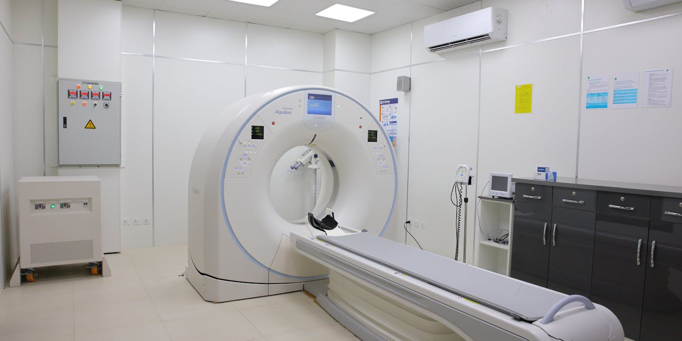
Spine MRI Report Explained
A plain-language guide to the words radiologists use — disc bulge, herniation, foraminal narrowing, stenosis, and more — by Dr. Waqas Mehdi, Neurosurgeon, Lahore
You Just Got Your MRI Report — What Do These Words Mean?
If you have recently had a spine MRI, you have probably read through the radiologist's report and felt a jolt of anxiety at phrases like "disc bulge," "foraminal narrowing," "facet arthropathy," or "degenerative changes." This reaction is completely normal — radiology reports are written by radiologists for other doctors, in a dense, technical shorthand, describing anatomy in exhaustive detail without any explanation of what any of it actually means for you, or how worried you should be.
Here is the reassuring truth: the vast majority of terms that appear on a spine MRI report describe changes that are common, often age-related, and frequently found in people who have no back pain at all. A report full of technical language does not automatically mean something is seriously wrong, and it does not automatically mean you need surgery. What it means is that the report needs to be correctly translated and then matched against your actual symptoms.
This page walks through the terminology that appears most often on lumbar and cervical MRI reports — in plain language — so that when you sit down with Dr. Waqas Mehdi, FCPS Neurosurgeon at MidCity Hospital Lahore, you already understand what the words mean and can focus the conversation on what matters most: what they mean for you specifically.
Understanding Vertebral Levels — What Do L4-L5 and L5-S1 Mean?
Your spine is divided into regions, and each vertebra (spinal bone) within a region is numbered from top to bottom. Radiologists describe findings by which two vertebrae — or which disc between them — is affected:
Cervical Spine (Neck) — C1 to C7
Seven vertebrae in the neck. A report might mention "C5-C6" or "C6-C7," the two most commonly affected cervical levels, referring to the disc between the fifth-sixth or sixth-seventh cervical vertebrae.
Lumbar Spine (Lower Back) — L1 to L5, then S1
Five lumbar vertebrae, followed by the sacrum (S1 and below). "L4-L5" means the disc between the fourth and fifth lumbar vertebrae; "L5-S1" means the disc between the fifth lumbar vertebra and the sacrum. These are, by a wide margin, the two most commonly affected — and most commonly searched — levels, because they bear the greatest mechanical load in the spine.
At each level, a specific nerve root exits the spine and travels down toward a particular strip of the leg or arm. As a general pattern (not an absolute rule — actual anatomy and compression location can vary):
Commonly produces pain or numbness along the outer thigh, outer calf, and top of the foot, and weakness in lifting the big toe or the foot upward (dorsiflexion) — sometimes noticed as the foot "slapping" or catching while walking.
Commonly produces pain radiating down the back of the thigh and calf into the outer edge of the foot and little toe, weakness pushing the foot downward (as when standing on tiptoe), and a reduced or absent ankle reflex.
These are typical patterns, not rigid rules — nerve anatomy varies slightly between individuals, and a compressive lesion can sometimes affect an adjacent root. This is exactly why Dr. Waqas Mehdi always correlates the level shown on your MRI with a hands-on examination of reflexes, strength, and sensation before drawing conclusions.
Disc Bulge vs Protrusion vs Extrusion vs Sequestration
These four words describe increasing severity or type along one single underlying process — a disc's outer wall being placed under strain — not four different diseases. Seeing one of these terms on your report simply tells you where, on this spectrum, your disc currently sits.
| Term | What's Happening | Typical Severity |
|---|---|---|
| Disc Bulge | Outer wall (annulus) intact but stretched outward broadly, around most of its circumference. | Mildest — often incidental, frequently symptom-free |
| Disc Protrusion | A more focal outward bulge in one direction; annulus fibres thinned but still contained. | Mild-moderate — the base is wider than the outward extension |
| Disc Extrusion | Inner nucleus breaks fully through the torn annulus wall, though it usually stays connected to the parent disc. | Moderate-severe — a true herniation, more likely to compress a nerve root directly |
| Disc Sequestration | A fragment of extruded material breaks off entirely and migrates within the spinal canal, separate from the parent disc. | Most severe by appearance — though free fragments are often reabsorbed by the body faster than contained extrusions |
For a deeper look at what causes disc herniation, why it produces leg or arm pain, and how each stage is actually treated, see the full guide: Herniated Disc — Causes, Symptoms & Treatment.
Foraminal Narrowing / Foraminal Stenosis
On either side of each vertebral level, there is a small bony tunnel called the neural foramen through which a single nerve root exits the spine on its way to the arm or leg. "Foraminal narrowing" or "foraminal stenosis" means this specific opening has become tighter than normal, usually from a combination of a bulging or herniated disc from in front, and enlarged facet joints or bone spurs from behind.
Because the foramen carries only one nerve root, foraminal narrowing tends to produce symptoms in a fairly specific, well-defined strip of the arm or leg corresponding to that root — rather than the more widespread pattern seen with central canal narrowing, described next. Mild foraminal narrowing without matching symptoms is common and, again, does not by itself require treatment.
Central Canal Stenosis
The central canal is the main bony tunnel running down the middle of the spine, carrying the spinal cord (in the neck and upper back) or the bundle of nerve roots called the cauda equina (in the lower back). "Central canal stenosis" means this main tunnel itself has narrowed — typically from a combination of disc bulging, thickened ligament, and facet joint enlargement pressing in from multiple sides at once, rather than a single nerve root being pinched.
Because it can affect multiple nerve roots or the spinal cord together, central canal stenosis tends to cause broader, less localized symptoms — such as both legs feeling heavy with walking (neurogenic claudication) in the lower back, or hand clumsiness and gait imbalance if it occurs in the neck. For the full picture on this condition, including how mild, moderate, and severe stenosis are actually managed, see Spinal Stenosis — Causes, Symptoms & Treatment.
Nerve Root Compression / Impingement
"Nerve root compression" or "nerve root impingement" on a report means the radiologist can see, on the images, that a specific nerve root is being physically touched, flattened, or displaced by a disc, bone spur, or narrowed foramen — as opposed to that structure simply narrowing without visibly contacting the nerve.
This is one of the more clinically meaningful phrases on a report, because true compression is more likely to correlate with the pain, numbness, tingling, or weakness radiating down the specific arm or leg pattern that nerve supplies. Even so, the final judgment always combines this imaging description with your actual examination findings — reflexes, strength testing, and the exact distribution of your symptoms — rather than the imaging word alone.
Facet Arthropathy
The facet joints are small paired joints at the back of each vertebra that let the spine bend, twist, and extend. Like any joint in the body — the knee or hip, for example — they can develop osteoarthritic wear over the years: cartilage thinning, joint space narrowing, and bone spur formation. This is what "facet arthropathy" or "facet joint degeneration" describes.
Facet arthropathy is an almost universal finding on spine MRI from the 40s onward and, in most people, causes little or no pain by itself. It becomes clinically important mainly in two situations: when it is severe enough to contribute meaningfully to foraminal or central canal narrowing described above, or when a targeted clinical examination (sometimes combined with a diagnostic facet joint injection) identifies it specifically as the source of localized back pain. Seeing "facet arthropathy" on your report is, on its own, rarely a reason for concern.
Degenerative Changes / Degenerative Disc Disease
"Degenerative changes," "degenerative disc disease," or "spondylosis" on a report all refer to the normal, expected wear-and-tear a spine accumulates with age — discs losing water content and height, mild bulging, endplate changes, and the facet changes described above. Despite the word "disease" in the name, this is best understood as a description of an aging process rather than a diagnosis of an illness.
This point is genuinely important and well established in the medical literature: multiple large studies scanning people with absolutely no back pain have found disc degeneration, bulging, and even some disc herniations in a large proportion of them, with the frequency of these findings increasing steadily with age regardless of whether the person has ever had symptoms. In other words, a spine MRI showing "degenerative changes" in a 45- or 55-year-old is common and expected — it does not, by itself, explain your pain or indicate a need for treatment.
For more detail on this specific condition and how it is actually managed when it is contributing to symptoms, see Degenerative Disc Disease.
The Most Important Point on This Page
An MRI report describes anatomy — it does not, by itself, decide whether you need treatment. Correlating the report with your actual symptoms and a hands-on examination is what determines the right next step. This is exactly why Dr. Waqas Mehdi's approach across every spinal condition he treats starts with matching what the scan shows to what you actually feel, and reserves surgery for cases where it is genuinely needed — not for every line item on a radiology report.
What Should You Do With This Report?
Bring your MRI report — and, ideally, the scan images or disc themselves — to a consultation with Dr. Waqas Mehdi, FCPS Neurosurgeon and Assistant Professor at King Edward Medical University (KEMU) and Mayo Hospital, Lahore. He will explain, in plain language, exactly what your specific report means, examine you to see whether the findings match your symptoms, and recommend the appropriate next step — which, for the large majority of patients, is conservative treatment rather than surgery.
If you already have a surgical recommendation based on your MRI and want an independent read before proceeding, a formal second opinion consultation is available.
Frequently Asked Questions
What does L4-L5 disc bulge mean?
Is a disc protrusion serious?
What is the difference between disc bulge and disc herniation?
Does everyone with degenerative changes on MRI need treatment?
What is foraminal narrowing?
Should I be worried about facet arthropathy on my MRI?
Can I get a second opinion on my MRI report?
Why does my MRI report look so much worse than how I actually feel?
Get Your MRI Report Explained by an Expert
Don't let confusing terminology cause unnecessary worry. Bring your MRI report and scans to Dr. Waqas Mehdi for a clear, honest explanation of what it means for you.
Consult Dr. Waqas Mehdi Today
Pakistan's trusted neurosurgeon for brain tumor surgery, spine surgery, and all complex neurological conditions. Book your appointment at MidCity Hospital, Lahore.